


What is electromyography and how does it help you?
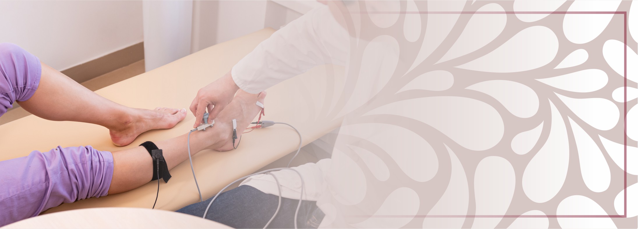
Electromyography (EMG) is a procedure that assesses nerve and muscle function. This examination is essential in establishing an accurate diagnosis of the pathology of the peripheral nervous system.
The terms electromyogram or electromyography are currently used for the entire electrodiagnostic evaluation, which includes nerve conduction studies and needle electromyography.
Content
- When is electromyography recommended?
- What is the electromyographic examination (EMG)?
- Are there any risks or contraindications?
- Anaesthesia and EMG examination
When is electromyography recommended?
An electrodiagnostic test is indicated in the evaluation of the function of the peripheral nervous system, of the neuromuscular junction and the muscular activity.
It is useful in diagnosing the following types of conditions:
Peripheral nerve disorders:
- hereditary neuropathies - Charcot Marie Tooth disease,
- acquired neuropathies
- posttraumatic: ulnar, median, radial nerve mononeuropathy after fractures; incarceration injuries ex. carpal tunnel syndrome,
- inflammatory/autoimmune - acute polyradiculoneuritis (Guillain Barre SDR), chronic demyelinating polyradiculoneuritis (CIDP), multiplex neuritis, etc.
- brachial plexus disorders:
- brachial plexus obstetric paralysis from the age of the newborn / infant;
- post-traumatic brachial plexus paralysis after road accidents
Anterior horn disease (spinal cord injury):
- degenerative type - spinal amyotrophy
- acquired - myelitis, neuroviruses, spinal cord injuries, vascular disorders with motor damage to the spinal neuron
Neuro-muscular junction diseases using special techniques - repetitive stimulation (SR) or EMG on single muscle fibre (SF EMG)
- myasthenia gravis
- congenital myasthenic syndromes
- botulism
Muscle diseases:
- congenital myopathies, inflammatory myopathies (dermatomyositis, postviral myositis) muscular dystrophies, mitochondrial myopathies
Other indications for EMG using special techniques
- evaluation of cranial nerves (V, VII, XII), evaluation of swallowing disorders of neurological cause, channelopathies - myotonic / non-myotonic dystrophies, periodic paralysis
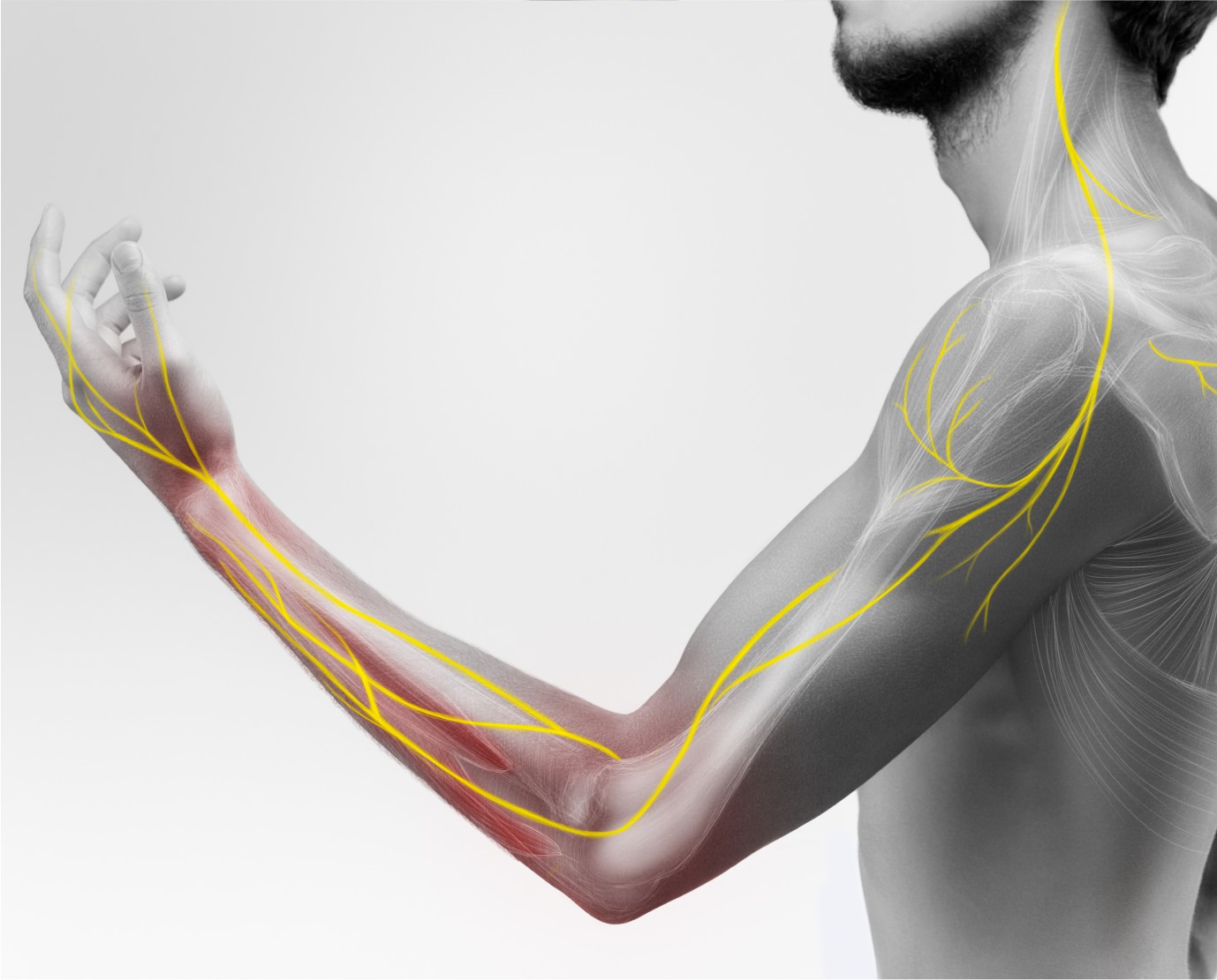 Electrodiagnostic testing is the only way to assess the functionality of the peripheral nervous system.
This helps the specialist in locating the peripheral nervous system/muscle condition, in assessing the type of peripheral nerve damage (axonal or demyelinating) and its severity.
It also provides the surgeon with information on the indication for surgery on the peripheral nerve or spine (herniated discs with severe radiculopathy diagnosed electromyographically), an indication that cannot always be established based on imaging examinations.
Electromyography can be recommended by a medical neurologist, neurosurgeon, physical medicine and recovery doctor, orthopedist, rheumatologist, diabetologist, internist, plastic surgeon.
What is the electromyographic examination (EMG)?
The electromyographic examination consists of two parts. In special situations, the doctor may opt for only one of these. Both sides of the test are well tolerated by most patients, although they cause discomfort and do not pose a significant risk to subsequent health.
The first part of the procedure is the study of nerve conduction. Electronurography involves stimulating the nerves at various points along their path through a small electrical discharge. The doctor will apply small electrodes to the skin for recording, usually on the hand and foot, and will apply a small electric shock to the skin in another area of the limb where the recording is being made. The sensation that the patient will perceive may or may not be painful.
The second part of the procedure is the needle muscle examination - the electromyography itself - is performed by inserting a very thin needle into certain muscles. The needle contains a microscopic electrode that records the normal or abnormal electrical activity of the muscle fibres. The needle is inserted into the relaxed muscle and moved a little to record muscle activity at rest, then the doctor will ask the patient to tighten the studied muscle to assess the muscle while is active.
The less pleasant moment is the piercing of the skin with the needle, the movements of the needle in the muscles being less perceived as pain, but more as a sensation of pressure. The needle has only a recording role, through which no electric shocks or intramuscular injections are administered.
Are there any risks or contraindications?
There are no significant risks of electromyographic examination; the patient may experience small bruises at the puncture site in the muscles and discomfort during the examination.
After the EMG exam, the usual daily activity can be resumed. The patient can drive. Any discomfort after the examination should disappear completely within 24-48 hours.
There are no dietary restrictions before or after the test, the patient can come and go by car/can drive and can continue his normal activity for the rest of the day.
Do not apply any product on the skin on the day of examination (lotions, oils, creams).
It is recommended that the clothing be light because it is often necessary to examine areas such as the shoulder, thigh or back.
Medications that are prescribed can be taken with a few notable exceptions.
The patient should inform the doctor before the procedure if he is undergoing anticoagulant treatment, as in this case, the needle examination may lead to bleeding inside the muscles. If needle examination is necessary, then oral anticoagulant treatment should be discontinued with the advice of the attending physician.
Also, if the patient is being treated for Myasthenia Gravis, the anticholinesterase medication (Myostin, Mestinon) should be stopped 48 hours in advance, with the advice of the attending physician.
Patients who have a blood clotting condition (severe liver disease, severe thrombocytopenia, haemophilia, etc.) may not have a needle EMG.
There are no contraindications to the study of nerve conduction.
Anaesthesia and EMG examination
Throughout the examination, the patient must cooperate as completely as possible. The discomfort caused during the EMG examination is perceived by most patients as minor and does not justify the anaesthetic risks. For these reasons, the EMG examination is performed without anaesthesia.
The article was written by Dr Ionela Codita - Primary Care Neurologist